43 label diagram of microscope
Parts of the Microscope with Labeling (also Free Printouts) 5. Knobs (fine and coarse) By adjusting the knob, you can adjust the focus of the microscope. The majority of the microscope models today have the knobs mounted on the same part of the device. Image 5: The circled parts of the microscope are the fine and coarse adjustment knobs. Picture Source: bp.blogspot.com. Labeling the Parts of the Microscope Labeling the Parts of the Microscope This activity has been designed for use in homes and schools. Each microscope layout (both blank and the version with answers) are available as PDF downloads. You can view a more in-depth review of each part of the microscope here. Download the Label the Parts of the Microscope PDF printable version here.
Labeled Diagram Of A Stereo Microscope | Products & Suppliers ... Products/Services for Labeled Diagram Of A Stereo Microscope. Microscopes - (705 companies) Microscopes are instruments that produce magnified images of small objects Microscopes are instruments that produce a magnified image of a small object. They are used in many scientific and industrial applications.

Label diagram of microscope
22 Parts Of a Microscope With Their Function And Labeled Diagram The field diaphragm control is located around the lens located in the base. Hinge Screw -This screw fixes the arm to the base and allow for the tilting of the arm. Stage Clips - They hold the slide firmly onto the stage. On/OFF Switch - This switch on the base of the microscope turns the illuminator off and on. Microscope Parts and Functions With Labeled Diagram and ... Most specimens are mounted on slides, flat rectangles of thin glass. The specimen is placed on the glass and a cover slip is placed over the specimen. This allows the slide to be easily inserted or removed from the microscope. It also allows the specimen to be labeled, transported, and stored without damage. PDF Label parts of the Microscope Label parts of the Microscope: . Created Date: 20150715115425Z
Label diagram of microscope. Microscope Labeling - The Biology Corner Students label the parts of the microscope in this photo of a basic laboratory light microscope. Can be used for practice or as a quiz. ... Microscope Labeling . Microscope Use: 15. When focusing a specimen, you should always start with the _____ objective. 16. When using the high power objective, only the _____ knob should be used. 17. The ... Microscope Types (with labeled diagrams) and Functions Dec 26, 2021 · Simple microscope labeled diagram Simple microscope functions It is used in industrial applications like: Watchmakers to assemble watches Cloth industry to count the number of threads or fibers in a cloth Jewelers to examine the finer parts of jewelry Miniature artists to examine and build their work Also used to inspect finer details on products Microscope Label Diagram | Quizlet Start studying Microscope Label. Learn vocabulary, terms, and more with flashcards, games, and other study tools. Parts of a microscope with functions and labeled diagram Head - This is also known as the body. It carries the optical parts in the upper part of the microscope. Base - It acts as microscopes support. It also carries microscopic illuminators. Arms - This is the part connecting the base and to the head and the eyepiece tube to the base of the microscope.
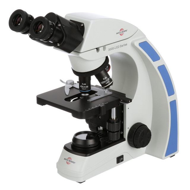
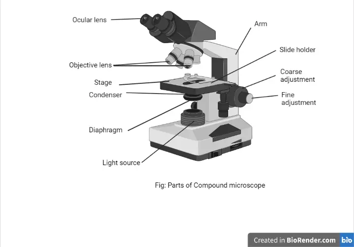
Microscope Labeling Diagram | Quizlet Unit 2 Lesson 5 - Punnett Squares and Pedigrees. 4 terms. PGFry210. Unit 2 Lesson 4 - Heredity. 9 terms. PGFry210. Upgrade to remove ads. Only $2.99/month. Label Microscope Diagram - EnchantedLearning.com Using the terms listed below, label the microscope diagram. arm - this attaches the eyepiece and body tube to the base. base - this supports the microscope. body tube - the tube that supports the eyepiece. coarse focus adjustment - a knob that makes large adjustments to the focus. diaphragm - an adjustable opening under the stage, allowing ... Microscope, Microscope Parts, Labeled Diagram, and Functions Revolving Nosepiece or Turret: Turret is the part of the microscope that holds two or multiple objective lenses and helps to rotate objective lenses and also helps to easily change power. Objective Lenses: Three are 3 or 4 objective lenses on a microscope. The objective lenses almost always consist of 4x, 10x, 40x and 100x powers. The most common eyepiece lens is 10x and when it coupled with ... 16 Parts of a Compound Microscope: Diagrams and Video Once you have an understanding of the parts of the microscope it will be much easier to navigate around and begin observing your specimen, which is the fun part! The 16 core parts of a compound microscope are: Head (Body) Arm. Base. Eyepiece. Eyepiece tube.
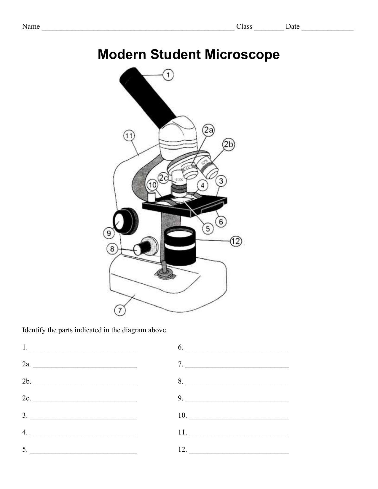
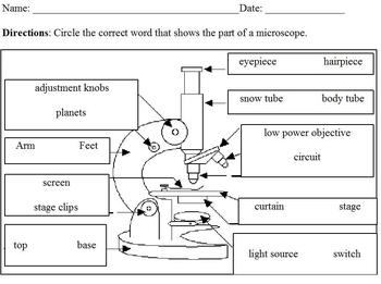
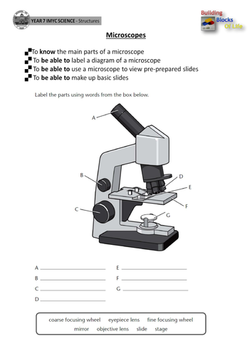
PDF Parts of a Microscope Printables - Homeschool Creations Label the parts of the microscope. You can use the word bank below to fill in the blanks or cut and paste the words at the bottom. Microscope Created by Jolanthe @ HomeschoolCreations.net. Parts of a eyepiece arm stageclips nosepiece focusing knobs illuminator stage objective lenses A Study of the Microscope and its Functions With a Labeled Diagram These labeled microscope diagrams and the functions of its various parts, attempt to simplify the microscope for you. However, as the saying goes, 'practice makes perfect', here is a blank compound microscope diagram and blank electron microscope diagram to label. Download the diagrams and practice labeling the different parts of these ... Compound Microscope Parts, Functions, and Labeled Diagram Compound Microscope Definitions for Labels. Eyepiece (ocular lens) with or without Pointer: The part that is looked through at the top of the compound microscope. Eyepieces typically have a magnification between 5x & 30x. Monocular or Binocular Head: Structural support that holds & connects the eyepieces to the objective lenses. Microscope labeled diagram - SlideShare Microscope labeled diagram 1. The Microscope Image courtesy of: Microscopehelp.com Basic rules to using the microscope 1. You should always carry a microscope with two hands, one on the arm and the other under the base. 2. You should always start on the lowest power objective lens and should always leave the microscope on the low power lens ...
Types of Microscopes: Definition, Working Principle, Diagram ...
A Study of the Microscope and its Functions With a Labeled Diagram Download Clker's Microscope With Labels clip art and related images now. Multiple sizes and related images are all free on Clker.com. A diagram showing all of the parts of a compound light microscope. Mar 7, 2021 - This Pin was discovered by Gloria Legg. Discover (and save!) your own Pins on Pinterest.
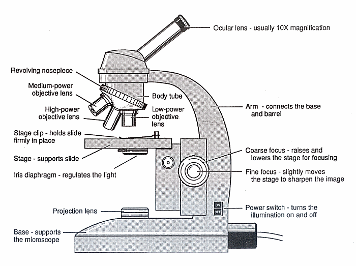
Diagram of a Microscope - Guide to using a microscope
Parts of Stereo Microscope (Dissecting microscope) - labeled diagram ... Labeled part diagram of a stereo microscope Major structural parts of a stereo microscope. There are three major structural parts of a stereo microscope. The viewing Head includes the upper part of the microscope, which houses the most critical optical components, including the eyepiece, objective lens, and light source of the microscope.

Parts of a Compound Microscope and Their Functions
Microscope Parts, Function, & Labeled Diagram - slidingmotion Objective lenses. Objective lenses are the most important part of the microscope. Its purpose is to visualize the specimen. There are 3-4 types of different objective lenses in any microscope. It has a magnification power of 4X to 100 X. 4X objective lens is the shortest lens while the 100X lens is the longest in terms of visualization.

Solved tration Questions: (10 points) Label the diagram of a ...
Label the Microscope Diagram | Download Scientific Diagram Download scientific diagram | Label the Microscope Diagram from publication: Laboratory Exercises in Microbiology: Discovering the Unseen World through Hands-on Investigation | Microbiology ...
Compound Microscope Parts, Functions, and Labeled Diagram ...
Labelled Diagram of Compound Microscope - Biology Discussion The below mentioned article provides a labelled diagram of compound microscope. Part # 1. The Stand: The stand is made up of a heavy foot which carries a curved inclinable limb or arm bearing the body tube. The foot is generally horse shoe-shaped structure (Fig. 2) which rests on table top or any other surface on which the microscope in kept.

Compound Microscope Parts – Labeled Diagram and their ...
Microscope Cell Labeled Under Leaf Search: Leaf Cell Under Microscope Labeled. After hundreds of years of observation, the cell theory was developed These leaves are two cells thick, so you should be able to focus up and down to see that the cells in one layer are larger than those in the other 8 Between the layers of cells inside the leaf are veins that contain xylem and After exposure to OGD for 4 h, followed by treatment ...
Microscope Parts and Function
Compound Microscope - Diagram (Parts labelled), Principle and Uses Image : Labeled Diagram of compound microscope parts. See: Labeled Diagram showing differences between compound and simple microscope parts Structural Components. The three structural components include. 1. Head. This is the upper part of the microscope that houses the optical parts. 2. Arm . This part connects the head with the base and ...

صلب وسط أسوأ الحد الأدنى غير راض من السهل حدوث ذلك a well labelled diagram of a microscope
Label the microscope — Science Learning Hub Jun 08, 2018 · Label the microscope Add to collection Use this interactive to identify and label the main parts of a microscope. Drag and drop the text labels onto the microscope diagram. eye piece lens coarse focus adjustment high-power objective diaphragm or iris base fine focus adjustment light source stage Download Exercise Tweet
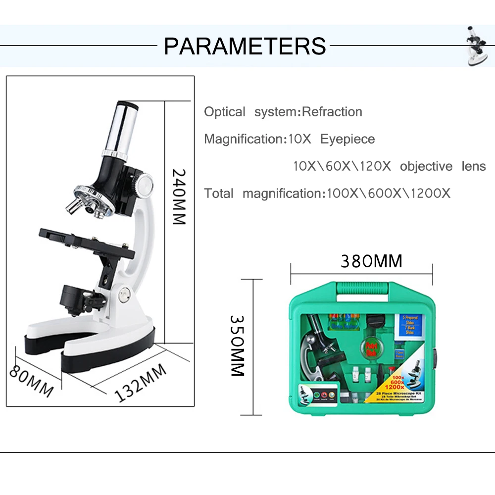
1200X Digital Microscope Set dengan Aksesoris Kit untuk Anak-anak Hadiah Siswa Semua Logam 100X 600X 1200X Putih Mikroskop
Compound Microscope Parts - Labeled Diagram and their Functions - Rs ... There are two major optical lens parts of a microscope: Eyepiece (10x) and Objective lenses (4x, 10x, 40x, 100x). Total magnification power is calculated by multiplying the magnification of the eyepiece and objective lens. The illuminator provides a source of light. The light is focused by the condenser and passing through the specimen placed ...

Diagram of traveling microscope setup with implant cast and ...
Parts of a Microscope Labeling Activity - Storyboard That Create a poster that labels the parts of a microscope and includes descriptions of what each part does. Click "Start Assignment". Use a landscape poster layout (large or small). Search for a diagram of a microscope. Using arrows and textables label each part of the microscope and describe its function.
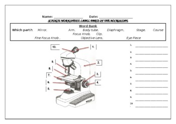
Science worksheet: Label The Parts Of A Microscope by Science ...
Microscope Labeling - The Biology Corner Microscope Labeling. Shannan Muskopf May 31, 2018. This simple worksheet pairs with a lesson on the light microscope, where beginning biology students learn the parts of the light microscope and the steps needed to focus a slide under high power. The labeling worksheet could be used as a quiz or as part of direct instruction where students ...

Microscope Diagram - Label Diagram | Quizlet
PDF Label parts of the Microscope Label parts of the Microscope: . Created Date: 20150715115425Z
Microscope - Teaching resources
Microscope Parts and Functions With Labeled Diagram and ... Most specimens are mounted on slides, flat rectangles of thin glass. The specimen is placed on the glass and a cover slip is placed over the specimen. This allows the slide to be easily inserted or removed from the microscope. It also allows the specimen to be labeled, transported, and stored without damage.

Microscope Labeling Diagram | Quizlet
22 Parts Of a Microscope With Their Function And Labeled Diagram The field diaphragm control is located around the lens located in the base. Hinge Screw -This screw fixes the arm to the base and allow for the tilting of the arm. Stage Clips - They hold the slide firmly onto the stage. On/OFF Switch - This switch on the base of the microscope turns the illuminator off and on.
Label a microscope - Teaching resources

Microscope parts 3D learning for Android - APK Download

Microscope Labeling
Parts of a Microscope with Their Functions • Microbe Online

eXe

Compound Microscope Parts, Functions, and Labeled Diagram ...
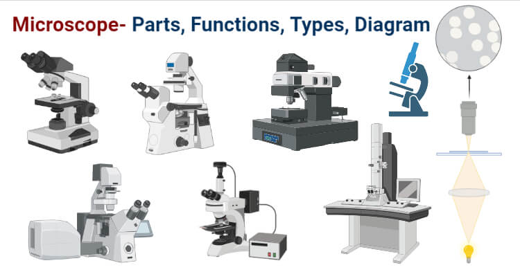
Microscope- Definition, Parts, Functions, Types, Diagram, Uses

How to draw Microscope diagram for beginners - step by step
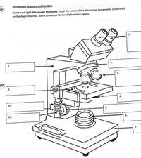
Answered: Microscope Structure and Function… | bartleby

Labeling the Parts of the Microscope | Microscope World Resources

Label Parts Of Microscope - ClipArt Best

Parts of Stereo Microscope (Dissecting microscope) – labeled ...

Parts of a Microscope Labeling Activity

Types of Microscopes: Definition, Working Principle, Diagram ...
Microscope labeling, modern and classical types

label microscope diagram | Charts | Microscope, Anatomy bones ...
Modified Science Diagram; Label Parts of a Microscope; Special Education

Microscope Parts and Functions

Draw a labelled diagram of a compound microscope.

BIOLOGY FROM 1 | EQUIPMENTS USED FOR OBSERVATION | Cours ...

How to draw compound of Microscope easily - step by step

Parts of a Microscope - SmartSchool Systems
Labeling Microscope Worksheet | Teaching Resources

Microscope Diagram Labeled, Unlabeled and Blank | Parts of a ...

5 Important Types of Microscopes used in Biology (With Diagram)
STEMI305 Stereo Microscope with integrated WIFI Camera Label ...

Microscope Diagram Labeled, Unlabeled and Blank | Parts of a ...

Living Environment Course

Parts of Microscope, Function, Names & Labeled Diagram ...
Post a Comment for "43 label diagram of microscope"